+91-9211592116

Call Support

1&2, Moti Ram Road, Ram Nagar

Shahdara, Delhi-110032

Home > ECG, EEG, HOLTER & EMG

ECG, EEG

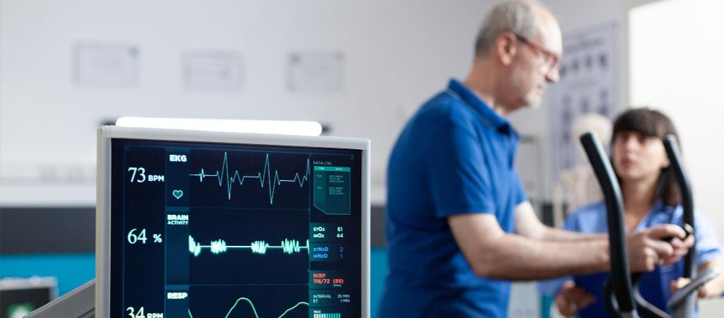
ECG (Electrocardiogram) and EEG (Electroencephalogram) are non-invasive diagnostic tools used to measure and record electrical activity in the body, primarily in the heart and brain, respectively. Each provides crucial information for diagnosing and monitoring various health conditions. Here’s a closer look at both:

ECG (Electrocardiogram)

What It Measures: The ECG measures the electrical activity of the heart. Each heartbeat generates electrical impulses, which the ECG captures as waveforms. These waveforms display the rhythm, rate, and strength of the heart’s activity.
How It Works: During an ECG test, electrodes are placed on specific locations on the skin of the chest, arms, and legs. These electrodes detect electrical signals produced by the heart’s movement and contractions. The ECG machine records these signals and produces a tracing on paper or a monitor.

Purpose and Applications:

  • Diagnosing heart conditions like arrhythmias, heart attacks, or heart muscle damage.
  • Monitoring heart health over time, especially after a heart surgery or when prescribed certain medications.
  • Evaluating symptoms like chest pain, palpitations, and dizziness.

EEG (Electroencephalogram)

What It Measures: The EEG captures and records electrical activity in the brain. Brain cells communicate through electrical impulses, which the EEG captures as waveforms or brain waves, representing different mental states.
How It Works: During an EEG test, multiple small electrodes are attached to the scalp. These electrodes record the brain’s electrical impulses and translate them into wave patterns. These patterns vary depending on the person’s level of consciousness, emotions, and cognitive activity.

Holter Monitor

A Holter monitor is a portable, continuous recording device that tracks electrical activity in the heart, much like an extended electrocardiogram (ECG or EKG). The purpose of the Holter monitor is to record heart rhythms over a prolonged period, typically 24-48 hours, although some may be worn for up to 7 days. This extended monitoring can capture irregular heartbeats or arrhythmias that might not appear in a short, standard ECG performed in a clinic.

  • How It Works: A Holter monitor is worn around the chest or waist, with electrodes attached to the skin to pick up the heart’s electrical signals. As the patient goes about their daily activities, the device records any variations in heart rhythms.
  • Uses: It’s especially helpful for diagnosing conditions like atrial fibrillation, palpitations, syncope (fainting), and unexplained chest pain. Holter monitors are also used to assess how well certain medications work, evaluate heart health after a heart attack, or monitor the heart’s response to treatment or pacemaker functions.
  • Benefits: The continuous monitoring over days provides a more comprehensive picture of heart function, especially if the symptoms are sporadic.
  • Limitations: Since Holter monitors are only worn temporarily, they may not capture very rare heart rhythm events. In such cases, an event monitor or implantable loop recorder may be considered instead.

Electromyography (EMG)

Electromyography (EMG) is a diagnostic procedure that assesses the health of muscles and the nerve cells that control them, known as motor neurons. EMG measures the electrical activity of muscles both at rest and during contraction. This helps determine whether muscle weakness or pain is due to a muscle issue or a nerve disorder.

  • How It Works: During an EMG, a fine needle electrode is inserted into the muscle tissue. The electrode detects electrical signals generated by muscle cells when they are activated by nerve impulses. These signals are then recorded and interpreted by a specialist.
  • Uses: EMG is used to diagnose conditions like carpal tunnel syndrome, amyotrophic lateral sclerosis (ALS), muscular dystrophy, nerve compression (like in radiculopathy), and other neuromuscular disorders. It’s helpful in assessing symptoms like muscle weakness, numbness, spasms, or unexplained pain.
  • Benefits: EMG can precisely detect nerve dysfunction, muscle dysfunction, or issues in nerve-to-muscle signal transmission. It helps doctors identify and locate nerve damage and determine the severity of a nerve or muscle disorder.
  • Limitations: EMG can be uncomfortable due to needle insertions, and interpreting the results requires specialized training. Additionally, EMG results are often combined with nerve conduction studies (NCS) for a complete picture.

Book Now