+91-9211592116
Call Support
1&2, Moti Ram Road, Ram Nagar
Shahdara, Delhi-110032
goodlifehealthcareorg@gmail.com
E-Mail Support
Call & Book Lab Visit
+91-9560428585/9560248585
Call Support
Shahdara, Delhi-110032
E-Mail Support
+91-9560428585/9560248585

X-rays: Often the first step in musculoskeletal imaging, X-rays are effective in detecting fractures, joint dislocations, and some degenerative conditions. They provide high-resolution images of bones but are less effective for soft tissue assessment.
MRI (Magnetic Resonance Imaging): MRI provides comprehensive soft tissue visualization, making it ideal for evaluating muscles, ligaments, cartilage, tendons, and other soft tissue structures. For instance, MRI is beneficial for identifying ligament tears in the knee or rotator cuff injuries in the shoulder. The detailed imaging helps to detect subtle injuries or degenerative changes.
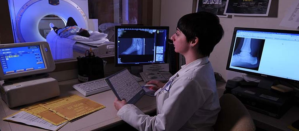
CT (Computed Tomography): CT scans are useful for providing cross-sectional images of bones and soft tissues, especially in complex areas like the hip or ankle. They are effective for detecting fractures, particularly in situations where bone alignment or joint structure needs to be assessed with precision.
Ultrasound: Ultrasound imaging is frequently used for evaluating superficial soft tissues, such as tendons and ligaments, and for guiding injections into joints. It’s especially beneficial for assessing injuries in the shoulder or ankle where ligaments and tendons are close to the surface.
Nuclear Medicine (Bone Scans): Bone scans can detect metabolic activity within bones, identifying areas of high activity, which may indicate fractures, infections, or tumors. This modality is useful for diagnosing unexplained pain when other imaging modalities do not provide conclusive results.
Shoulder: Commonly assessed for rotator cuff tears, labral injuries, and bursitis. MRI and ultrasound are particularly effective in diagnosing soft tissue injuries and assessing the extent of shoulder joint pathologies.
Knee: Knee imaging is often focused on assessing meniscal tears, ligament injuries (e.g., ACL or MCL tears), and cartilage damage. MRI is the preferred modality for its detail in soft tissue imaging, while X-rays are typically used to detect fractures or arthritis.
Wrist: Imaging of the wrist is essential for diagnosing fractures, ligament injuries, and conditions like carpal tunnel syndrome. MRI and ultrasound are useful for detailed soft tissue examination, and X-rays are used to detect fractures and joint alignment issues.
Ankle: Ankle imaging is commonly used for assessing ligament injuries, fractures, and tendonitis. MRI and ultrasound are frequently used for soft tissue injuries, while X-rays and CT scans are effective for bone fractures and alignment assessment.
Hip: Hip imaging often involves MRI and CT for identifying fractures, labral tears, and degenerative joint disease. These modalities provide excellent visualization of both bone and soft tissue structures, aiding in the diagnosis of complex hip pathologies.
Soft Tissue: Soft tissue imaging covers a range of structures, including muscles, tendons, ligaments, and bursae throughout the body. MRI and ultrasound are primary modalities here, allowing for a detailed examination of muscle tears, tendonitis, and soft tissue masses or infections.