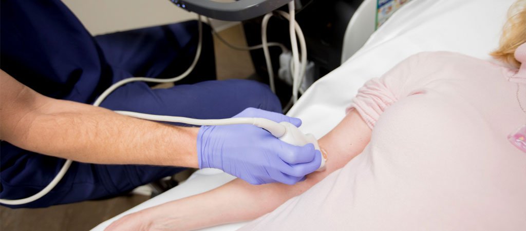
Small parts ultrasound is a specialized imaging technique used to examine various superficial structures in the body. It plays a critical role in evaluating localized swellings in regions such as the breast, scrotum, thyroid, chest, and eyes (B-scan). This non-invasive procedure provides real-time images, helping healthcare providers diagnose and manage various conditions effectively.
Breast ultrasound is commonly used to evaluate palpable lumps or areas of swelling. It helps differentiate between solid masses and fluid-filled cysts, guiding further management. This imaging modality is particularly beneficial for women with dense breast tissue, where mammograms may not provide clear results. The ultrasound can assist in determining the characteristics of a lump, such as its size, shape, and internal structure.
Scrotal ultrasound is used to assess localized swelling in the scrotum, which can indicate conditions like testicular torsion, varicocele, hydrocele, or tumors. This ultrasound helps visualize the testicles, epididymis, and surrounding structures, providing crucial information for diagnosis and treatment decisions.
Thyroid ultrasound is essential for evaluating nodules or swelling in the thyroid gland. It helps characterize the nodules based on their size, shape, echogenicity, and vascularity, aiding in the determination of whether further testing or intervention is necessary. This ultrasound is a critical tool in assessing thyroid disorders, including hyperthyroidism and hypothyroidism.
Chest ultrasound is used to evaluate localized swellings or abnormalities in the chest wall, pleura, and lung surfaces. It is particularly useful in assessing pleural effusions, lung masses, or other soft tissue abnormalities. This imaging technique provides real-time images, enabling the physician to guide procedures such as thoracentesis (draining fluid from the pleural space).
A B-scan ultrasound of the eye is performed to evaluate localized swelling or abnormalities within the eye that may not be visible during a standard eye examination. It helps diagnose conditions such as retinal detachment, tumors, and other intraocular disorders. The B-scan provides detailed images of the eye structures, allowing for accurate diagnosis and management.
